Diagnostic
Diagnostic Spectrum
Diagnostic spectrum of radiology
MRI:
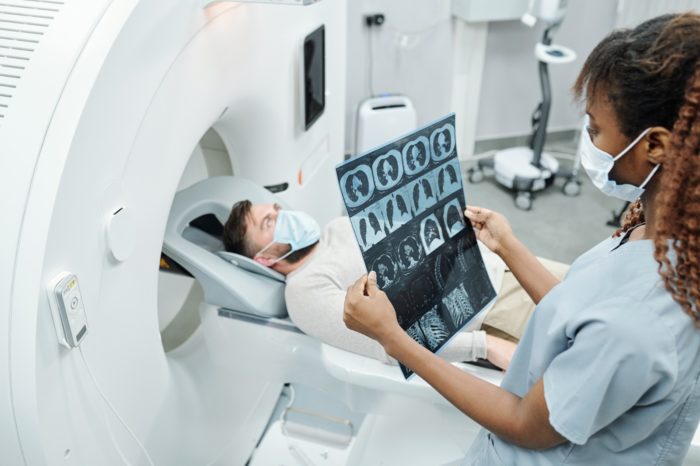
With MRT examinations, three-dimensional images can be created completely painlessly –without radiation exposure and in a layered procedure. MRI should not be confused with “X-rays” – or computed tomography. While X-rays are used to create images in these methods, the nuclear spin examination is based on a strong magnetic field within the “tube” in which the patient lies.
CT:
With this examination, it is also possible to visualize even the smallest structures. Tomography is an imaging procedure in radiology that is based on X-rays. The computer tomography sends X-rays through the body – and the body absorbs part of them. From these absorption values, the computer then calculates detailed sectional images, which – superimposed – can result in an overall picture

Nuclear-medicine:
In this examination procedure, a small amount of radioactive substance (radiopharmaceutical) is administered, which accumulates in the organs to be examined. There the radiopharmaceutical sends out weak radiation, which is detected by sensitive detectors. You can think of the device as a kind of camera that only records radioactive radiation. With the help of computers, an image (scintigram) is calculated from the recorded rays and this is assessed by the doctor on the screen. The method is suitable, for example, for locating foci of inflammation in the skeleton (skeletal scintigraphy) or for functional analysis of organs, for example in kidney function scintigraphy. The radiation exposure is comparable to that of a normal X-ray examination.
Ultrasound (sonography):
The ultrasound examination is an imaging procedure. It allows you to see into the human body from the outside. The internal organs can be viewed during sonography. Especially the soft tissues such as the liver, kidneys or spleen, which are difficult to see on X-rays. For the patient, sonography is comfortable, painless and without side effects.
Ultrasound diagnostics (sonography) is the most widely used imaging examination method in medicine, along with computed tomography and magnetic resonance tomography. This technique enables most of the structures of the internal body to be visualized by sending and receiving harmless ultrasonic waves. A gel is used for the examination because the sound waves cannot pass through the air and bones.
An ultrasound examination produces sectional images. With the Doppler method, the representation of motion sequences is possible. With this method it is possible to assess the function of the heart muscle, the intestines and joints.


