
RADIOLOGY
Radiology
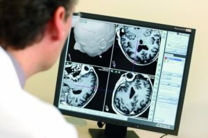
With magnetic resonance tomography, sectional images of the human body can be generated without X-rays. Radio waves are generated inside a spacious magnetic tube, which lead to reactions in the body, from which cross-sectional images of the examined body region can be created with the help of a computer.
Stroke, or temporary cerebral blood flow disorder of the brain (TIA: transistor Ischemic attack)
Cerebral hemorrhage, e.g .: subarachnoid hemorrhage (SAB), subdural and epidural hematoma, intracerebral hemorrhage (ICB)
– Vascular changes: vascular constrictions and occlusions in the head and neck area, vascular sacs (aneurysm) and malformations.
-Tumor in the central or peripheral nervous system: diagnostics, surgical planning, therapy monitoring, prognosis assessment -spinal changes, e.g .: disc changes, spinal canal stenosis, back pain, radiating pain Inflammation and infections, e.g .: multiple sclerosis, borreliosis, meningitis, encephalitis, liquor fistula, spondylodiscitis
-Degenerative diseases of the central nervous system, e.g. Alzheimer’s dementia, frontotemporal dementia (M. Pick), Parkinson’s syndrome, etc.
-Injuries and consequences of accidents: Changes in the brain, spine and spinal cord, especially for expert assessment
– Injuries to joints, cartilage structures, ligaments or tendon
-Disc prolapse
-Inflammatory diseases of the bone
-Inflammatory diseases of the organs
Abscesses (accumulations of pus in a normally non-existent body cavity) and fistulas (ducts connecting organs or from inside the body to the surface of the body)
-Spread of cancer or assessment of the course of cancer
-Alternative to computed tomography (CT) examination, because you are allergic to the contrast agent used or you are pregnant
Procedure and preparation before an MRI examination
An examination in the magnetic resonance tomograph usually takes between 20 and 40 minutes. You are pushed into a 60 cm (diameter) large and 160 cm long tube while lying down. If you suffer from claustrophobia, you can be given a sedative beforehand.
During the exam, you will hear loud knocking noises. These noises come from fast-switching electromagnets and unfortunately cannot be avoided. You will receive headphones or earplugs as protection.
You must lie still during the examination. Even small movements can lead to disturbances in the images. When examining the abdomen and chest, you sometimes have to hold your breath for a short time (10-20 seconds).
For the examination, it is important that you remove all metal (e.g. glasses, watch, jewelry). Small zippers and bra underwires can also lead to image disturbances. If you have a pacemaker, you must never go into an MRI scanner!
Contrast media significantly improve the informative value of an MRI and can even reveal crucial information. Contrast media containing gadolinium are mainly excreted via the kidneys.
We only use contrast media after a comprehensive consultation with the patient and after careful consideration of whether the use of a contrast agent really offers relevant additional information. Of course, we administer the smallest possible amount. Our team only uses contrast media that have been tested and approved as medicinal products in accordance with international and German guidelines.
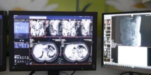
Computed tomography (CT) is an imaging process that uses a rotating, finely focused X-ray beam to generate high-resolution cross-sectional images of the inside of the body.
Computed tomography is a radiological examination procedure. In the computer tomograph, a system consisting of an X-ray tube or two X-ray tubes and a detector system with 16 – 128 lines circles the body in a spiral shape from which the computer calculates slice images.
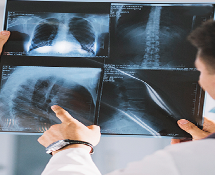
One of the classic areas of application of conventional X-rays is the chest image, which is still the most common examinations, whereby an assessment of the diaphragm, lungs, heart and the thoracic skeleton can be recognized. Another area of application of conventional X-rays is the examination of the skeleton, with bone fractures or pathological changes being diagnosed. In addition, the conventional X-ray can also be used to examine the abdominal region, in which, among other things, free air in the abdominal cavity or an intestinal obstruction can be detected. Mammography is also one of the conventional X-ray examinations, whereby a special X-ray device is required for the recording.
Before the examination, all external objects should be removed from the examination area (e.g. pieces of jewelry, bra hangers, etc.) in order to avoid overlapping. Special preparation is usually not necessary.
Depending on the specific question, X-rays are taken while standing, sitting or lying down. For the recordings, the X-rays are focused on the area of interest (e.g. hand, shoulder, thorax, etc.). Once the patient has been positioned appropriately for the desired perspective, an exposure can be taken in seconds. A fluoroscopic examination takes up to 30 minutes, depending on the question, and it may be necessary that you receive contrast media from us.